


Dermatological Research

Quantitative insight into skin pathophysiology and treatment efficacy
Non-invasive OCT imaging to visualise, measure and monitor dermatological disease — in real time
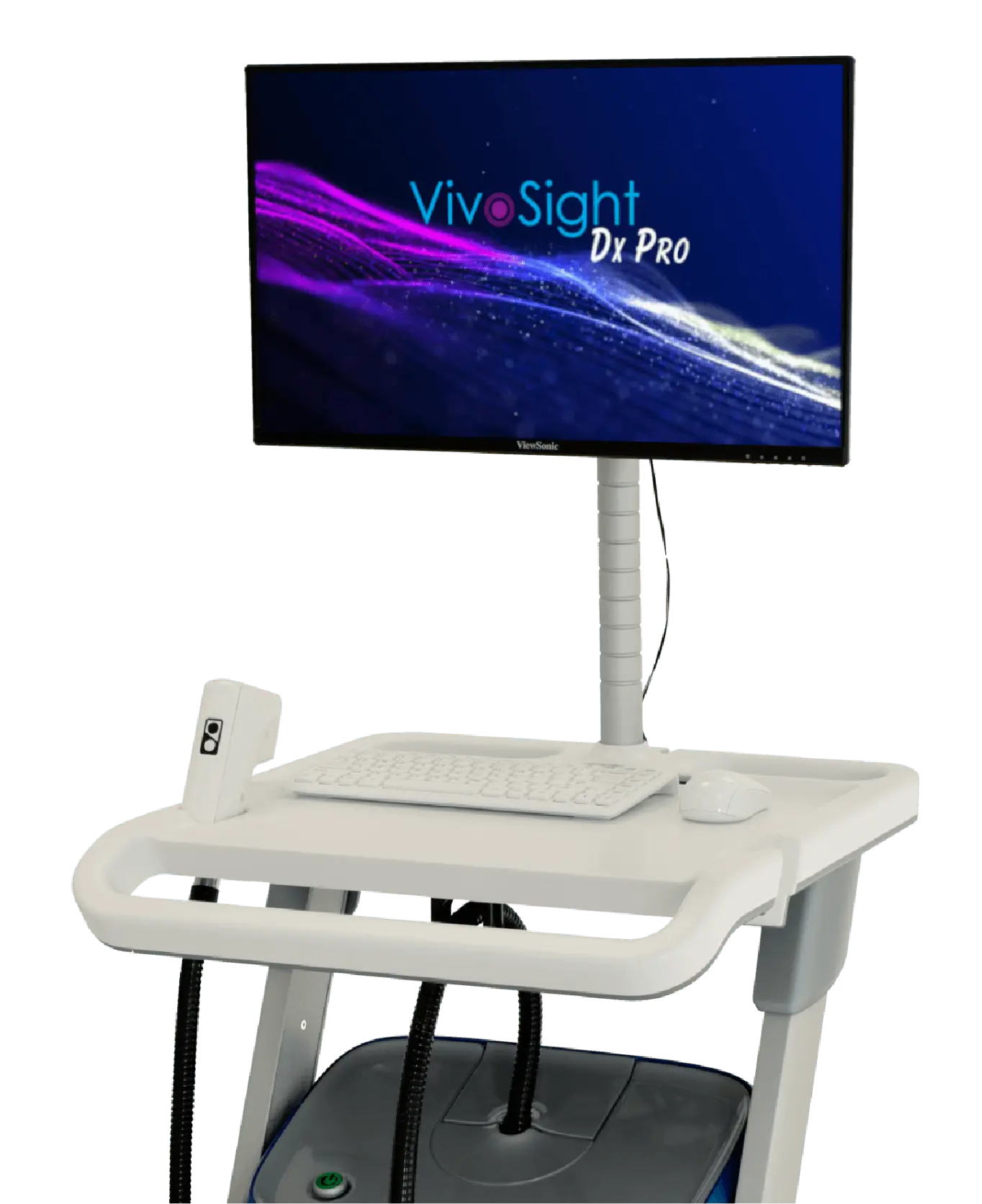
VivoSight Dx Pro delivers rapid, non-invasive and repeatable OCT imaging that enables objective assessment of skin structure, vascularity and treatment response — including sub-clinical effects.
Advance dermatological research
with objective data
VivoSight Dx Pro delivers high-resolution OCT imaging that enables quantitative, reproducible assessment of skin pathophysiology and therapeutic response, including sub-clinical effects. Imaging takes seconds and enables longitudinal monitoring with minimal burden on subjects.
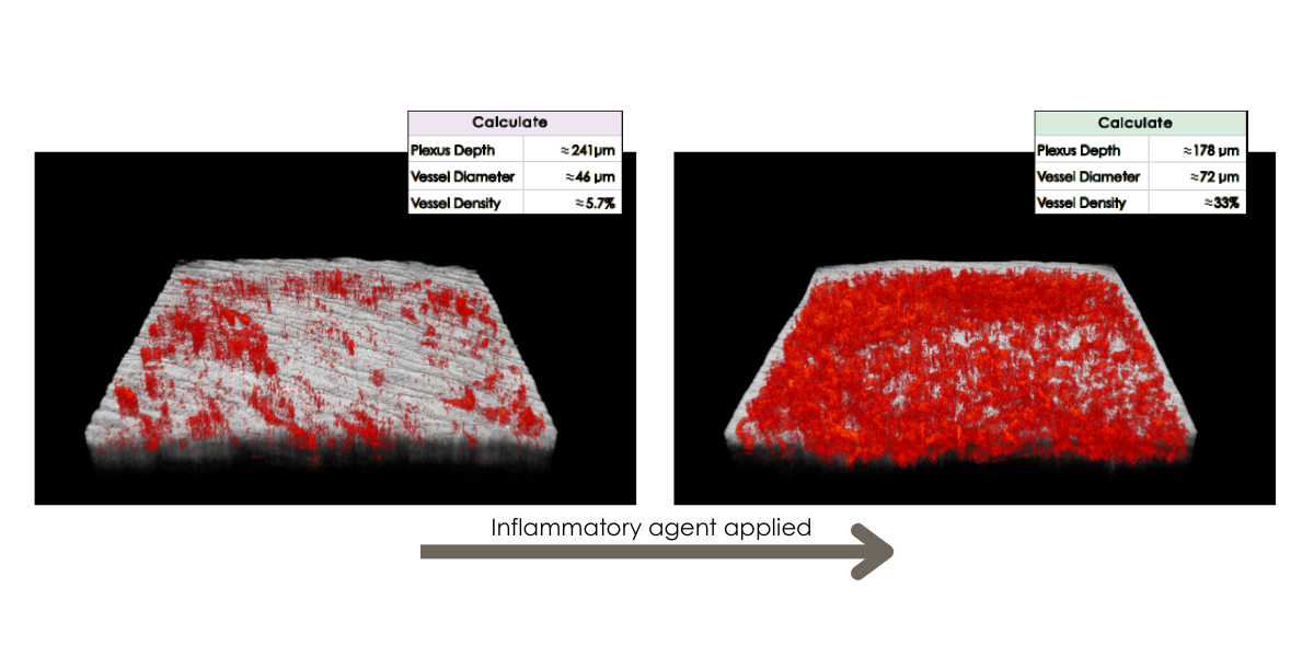
Quantify vascular changes.
Dynamic OCT enables visualisation and measurement of the superficial vascular plexus—supporting assessment of inflammation and angiogenesis over time.
Measure epidermal thickness with precision.
VivoTools extracts accurate, reproducible thickness measurements—ideal for evaluating treatments affecting skin barrier function.
Assess tissue density.
Quantitative analysis of epidermal density provides insight into fibrosis and collagen structure.
Generate objective, non-invasive endpoints.
High-resolution imaging combined with automated analysis supports consistent, repeatable research outcomes.
Strengthen study endpoints with non-invasive OCT
Quantify epidermal and vascular changes — including sub-clinical effects — across longitudinal studies.
Designed for clinical and translational research
- Device cleared for use in clinical settings
- Completely non-invasive — well accepted by subjects and patients
- Full 3D OCT scans acquired in just 15 seconds
- Repeatable measurements for longitudinal studies
- Comprehensive quantitative analysis to accelerate dermatological research
- Cost-effective compared with subjective qualitative measures
Fast, simple skin imaging
Quantify key disease and treatment parameters
VivoSight Dx Pro enables direct visualisation and objective measurement of structural and vascular changes associated with dermatological disease and therapy.
With the optional VivoTools® skin analysis software, researchers can quantify:
- Epidermal remodelling and thickness
- Alterations in vascular morphology
- Vessel diameter, density and depth as markers of inflammation
- Dermal brightness as a proxy for collagen density
- Skin surface roughness
These quantitative metrics support robust evaluation of disease severity, treatment response and sub-clinical effects.
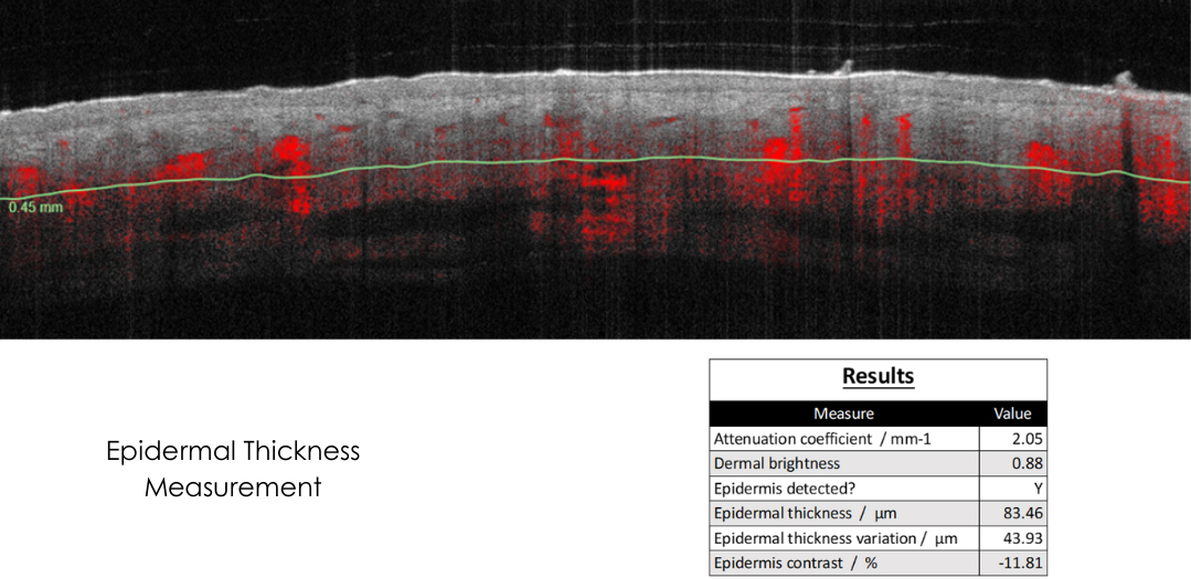
Multiple skin quality measures available, including Epidermal thickness
“OCT distinguishes micromorphology of the condition, and excellent vascular imaging capabilities add another layer to characterise pathology — often indicating degree of disease severity including sub-clinical disease.”
Prof. Giovanni Pellacani, MD, Chair of Dermatology, Sapienza University of Rome
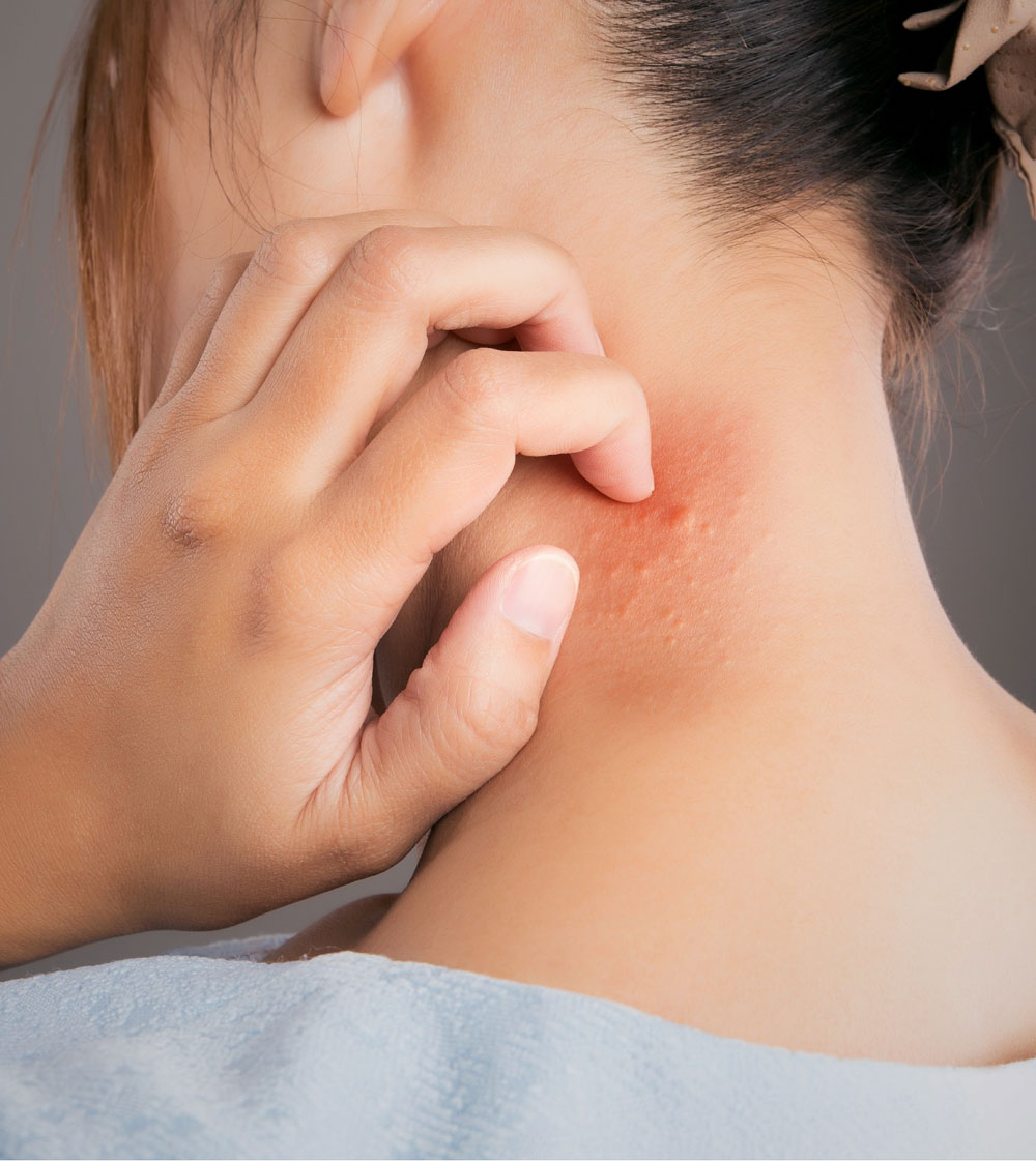
Study inflammatory and autoimmune skin disease
VivoSight Dx Pro has been used to assess a wide range of dermatological conditions, including:
Atopic dermatitis – Psoriasis – Acne – Systemic Sclerosis – Bullous diseases – Parasites – Alopecia – Onychomycosis & Nail diseases – Non-Melanoma & Melanoma Skin Cancer – Acute & Chronic Wound Healing – Scars – Port Wine Birth Marks & Vascular conditions – Diabetes – and many others
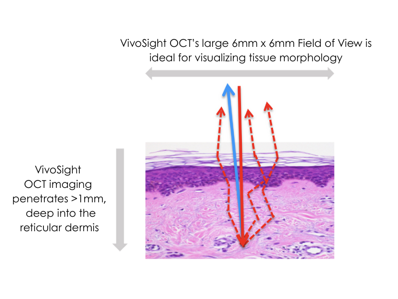
Wide 6 × 6 mm field-of-view imaging combined with >1 mm penetration depth provides detailed insight into epidermal structure and vascular networks, supporting deeper understanding of pathology and therapeutic impact.
Unrivalled visualisation
Transform dermatological
research with VivoSight
Reveal sub-clinical effects. Strengthen study endpoints. Accelerate discovery.
From early-stage pathophysiology studies to longitudinal assessment of therapeutic response, VivoSight Dx Pro provides objective, repeatable and non-invasive insight into skin disease — in real time.
VivoSight measurements are for investigational use only.