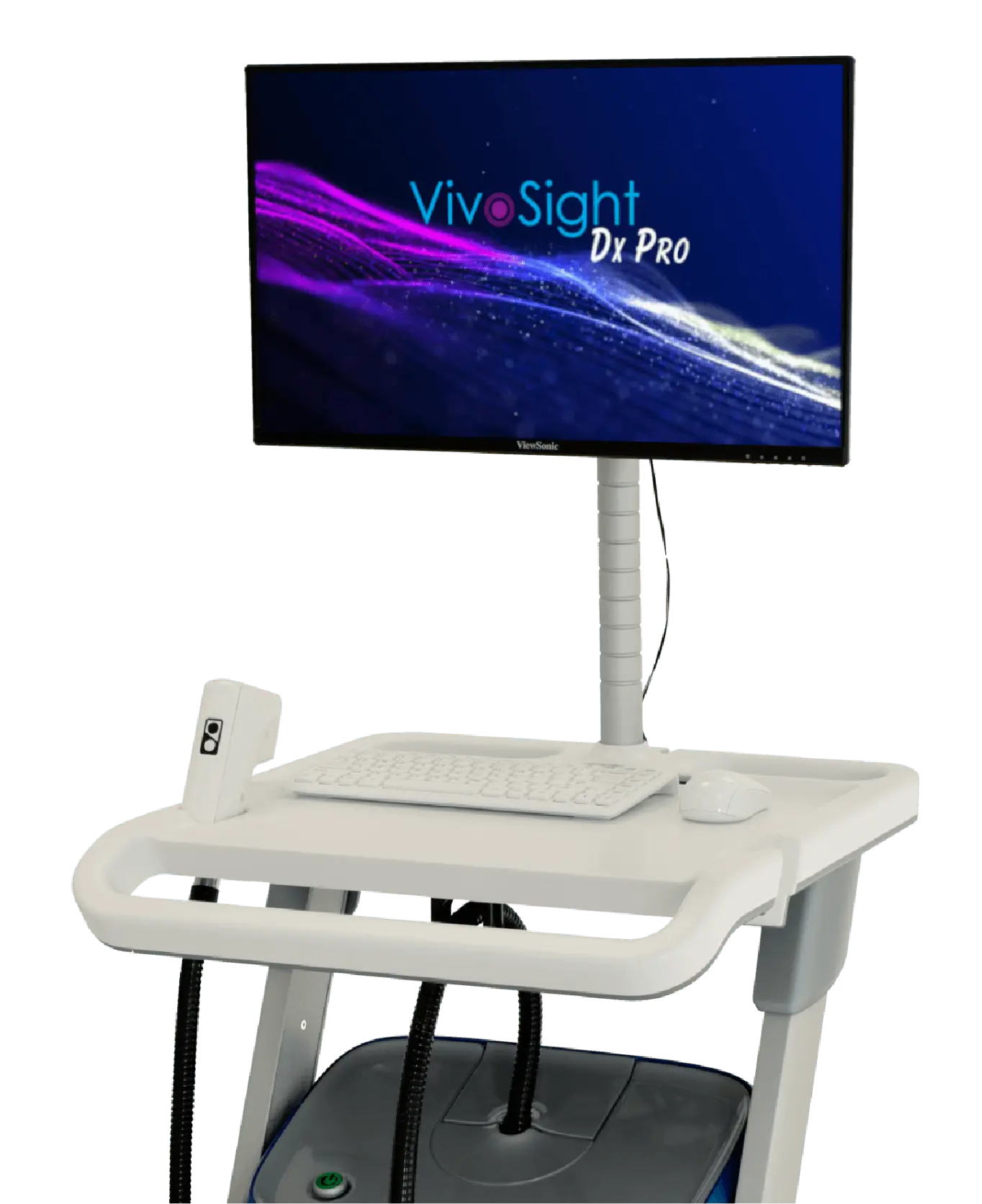
Skin Imaging – Reimagined
Faster, more confident, non-invasive skin assessment – without biopsy
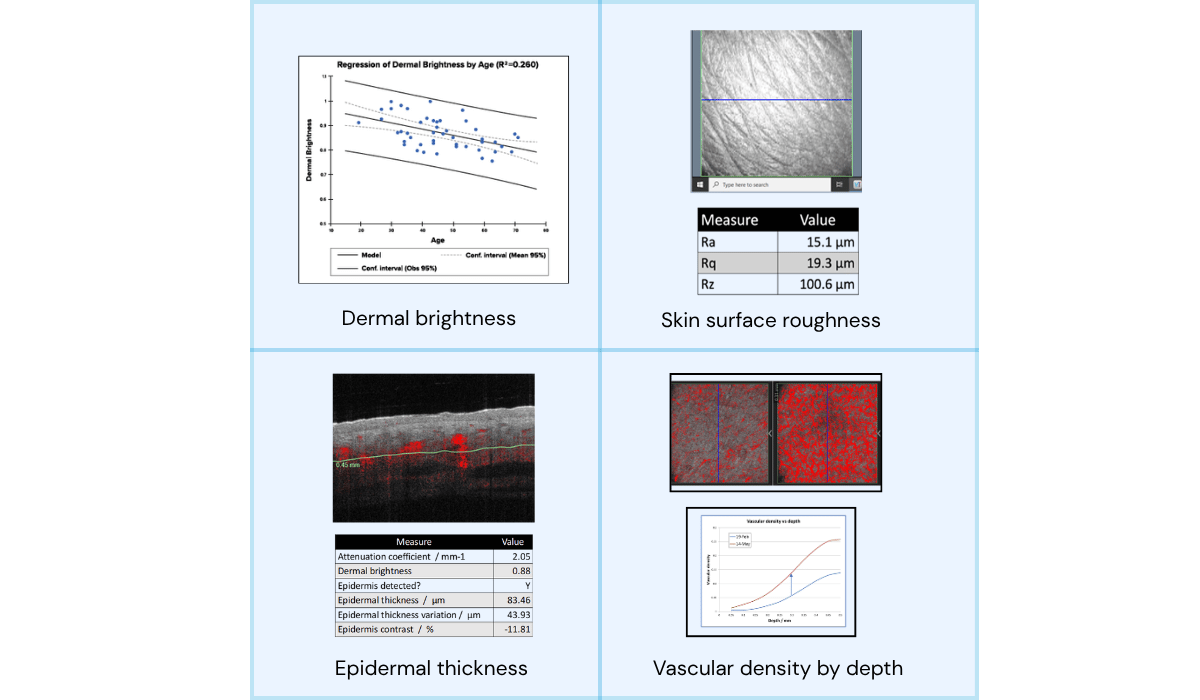
High-resolution, non-invasive OCT imaging that reveals
skin structure and microvasculature in 3D — in just seconds.
15-second scan • >1 mm imaging depth • 6 × 6 mm field of view • AI-assisted interpretation
VivoSight gives you
The Whole Picture®
VivoSight provides fast, non-invasive visualisation of skin structure and microvasculature, supporting confident assessment in both clinical practice and skin research.
-

Confident clinical decisions
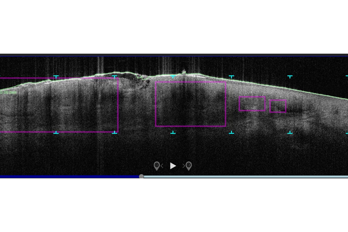
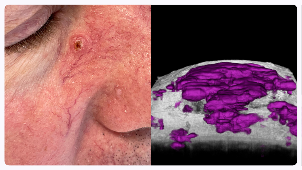
Clinically validated sensitivity and specificity support confident diagnosis, treatment selection and monitoring of Basal Cell Carcinoma (BCC) and other non-melanoma skin cancers.
With >1 mm imaging depth and a wide 6 × 6 mm field of view, VivoSight enables full visualisation of lesion architecture rather than partial sampling.
-

Efficient
workflowsA complete, high-resolution 3D scan is
acquired in just 15 seconds, capturing
the entire lesion in a single non-invasive
scan. -

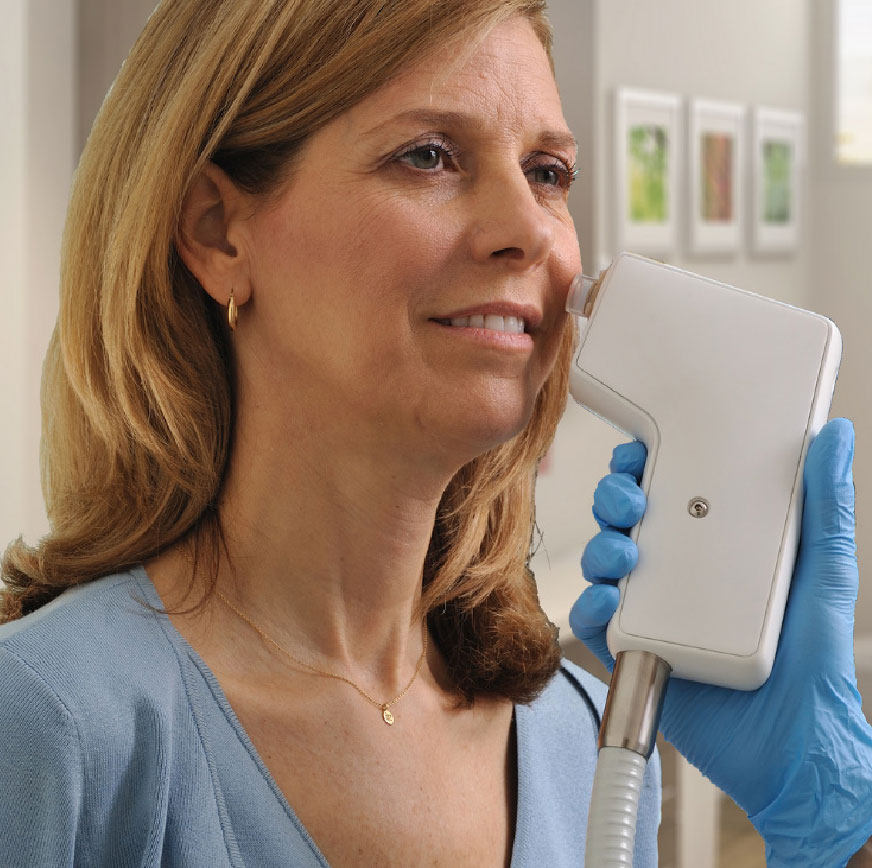
Better patient and subject experience
Completely non-invasive, scar- and pain-
free imaging will delight patients and enable
repeat assessment over time.
Interested? Schedule a Demo Today!
Our team is available for your online demo of VivoSight skin imaging. Complete the form in the link below to schedule a time with one of our experts.

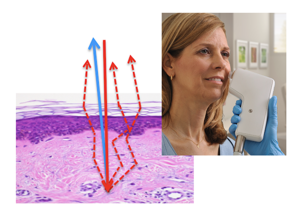
How OCT skin
imaging works
Optical Coherence Tomography (OCT) is an optical analogue of ultrasound imaging.
Using eye-safe infrared light, VivoSight captures a high-resolution 3D block of image data, revealing skin structure and function beneath the surface. This enables real-time, in-clinic visualisation of skin architecture without cutting, scarring, or waiting for histology.
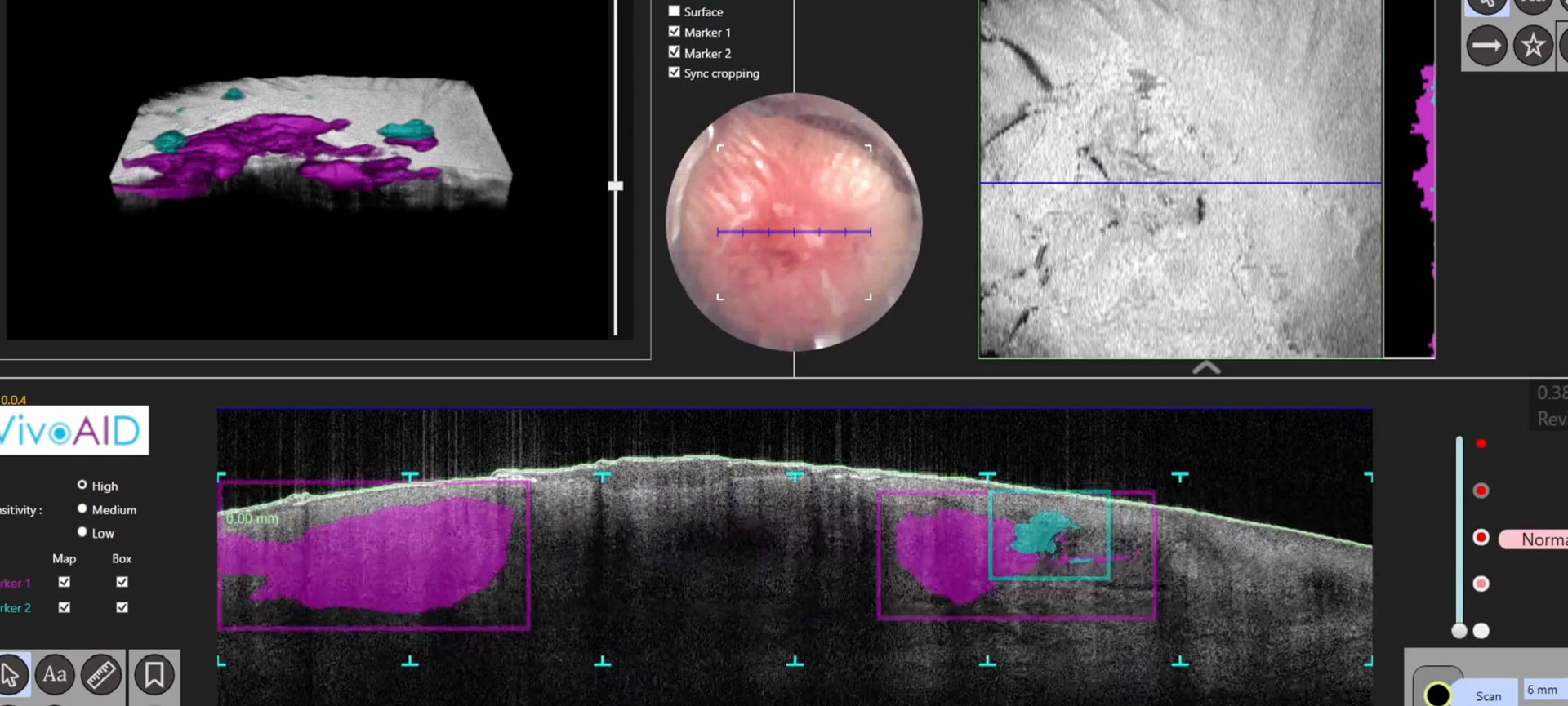
AI supporting clinical expertise
AI-assisted image analysis highlights clinically relevant features within OCT scans, helping clinicians interpret complex data more quickly and consistently. All decisions remain firmly clinician-based.
Explore AI-Assisted Imaging.
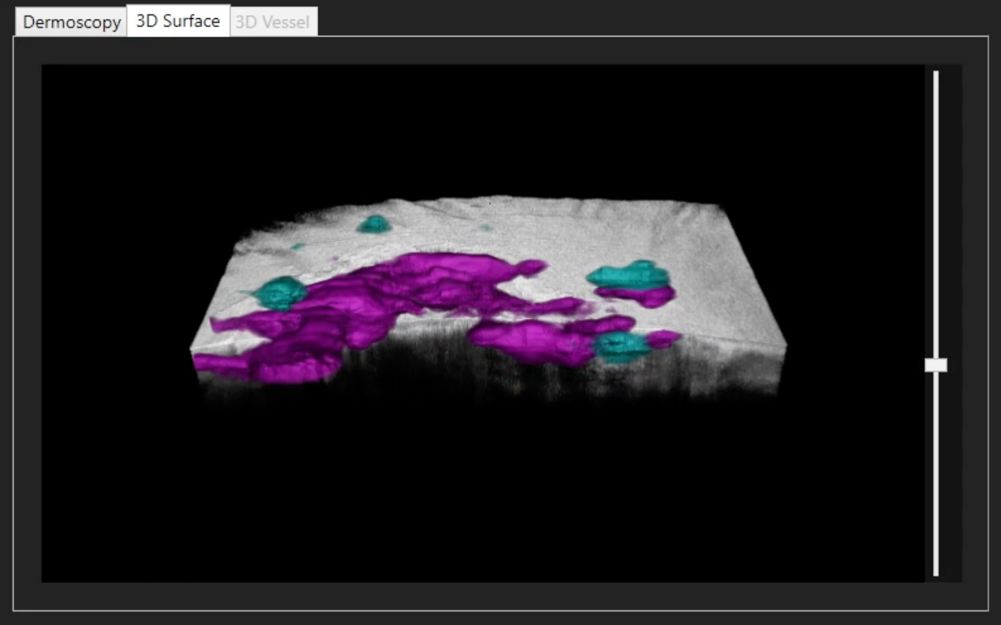
Real-time 3D rendering aids lesion visualization
Automatic placement of BCC image markers can speed clinician interpretation
Applications across dermatology and skin research
-

Clinical Dermatology
Supporting diagnosis, treatment selection, and monitoring across a wide range of non-melanoma skin conditions, integrating seamlessly into clinical workflows
-

Skin Research
Enable non-invasive, repeatable imaging for academic and commercial skin health research.
Trusted by clinicians and
researchers worldwide
Makes OCT skin imaging accessible, faster,
and easier to use.
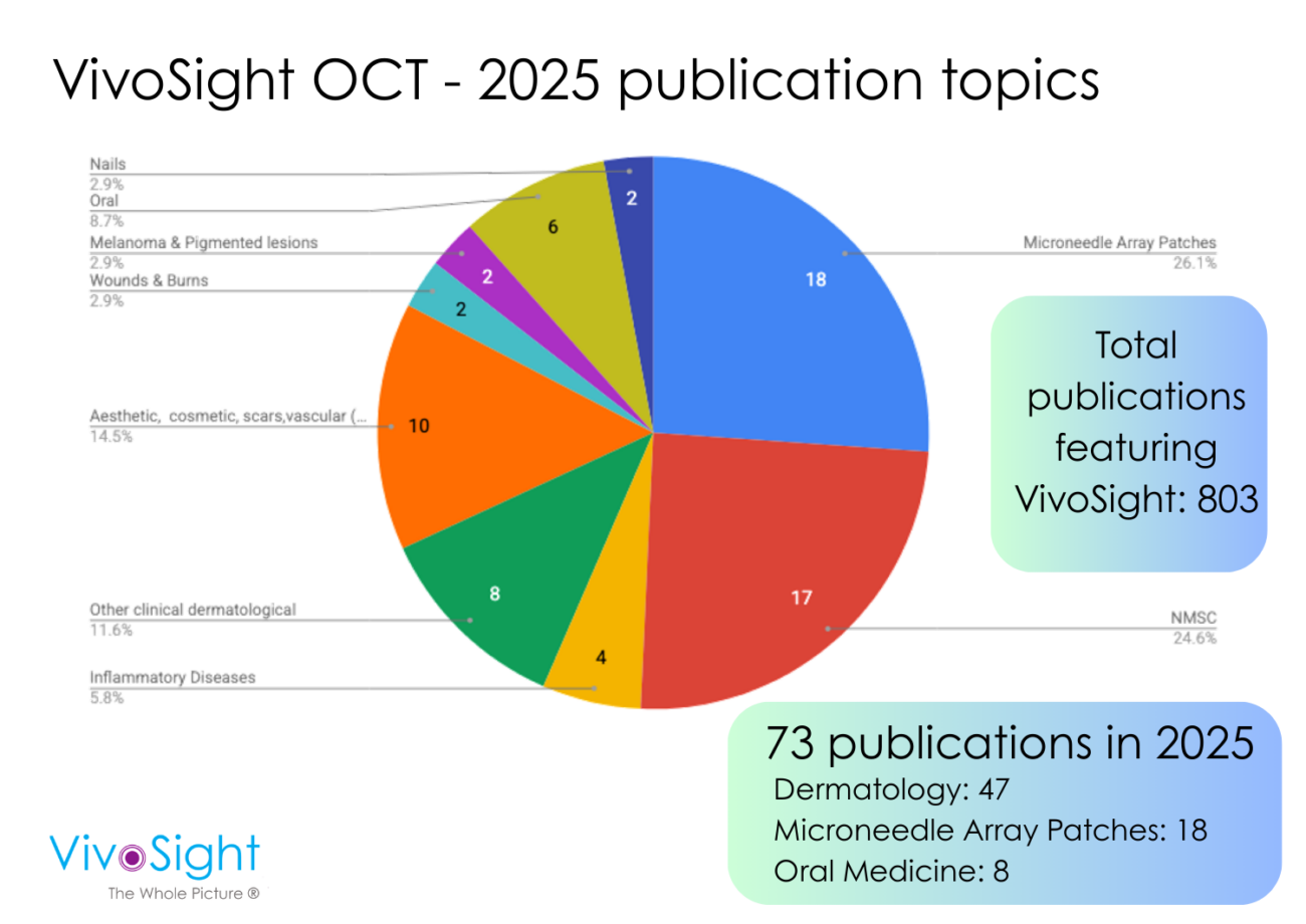
- 800+ peer-reviewed publications
- 200,000+ patient scans completed
- Used routinely in hospitals, clinics and research labs globally
- EU MDR certified
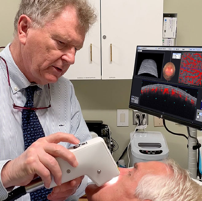
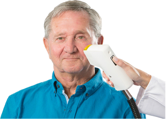
Prof. C. Zachary, UCI, using VivoSight to examine a patient
News & Insights
See VivoSight in Practice
Tailored to your clinical or research needs
Book an online demo with our team to see how VivoSight can support your clinical or research workflow.