


Non-Melanoma Skin Cancer (NMSC)
Diagnosis & Follow-Up

Faster, non-invasive insight
for confident BCC diagnosis
OCT skin imaging to simplify diagnosis, follow-up and clinical workflow.
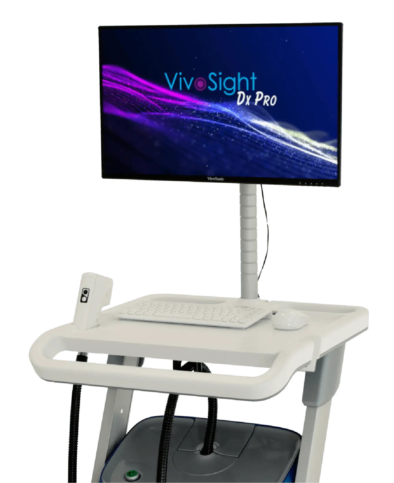
VivoSight Dx Pro delivers rapid, non-invasive OCT imaging that supports accurate BCC diagnosis and treatment follow-up — without routine biopsy.
OCT imaging is now Included in the European Consensus Guidelines for Diagnosis & Treatment of BCC, SCC & AK; and in many national Guidelines, including those of Germany and The Netherlands.
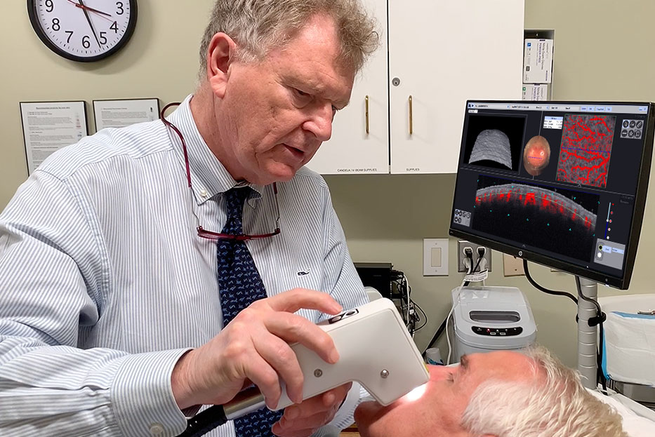
Professor Paasch, a practising dermatologist in Germany, explains how VivoSight OCT and VivoAID support confident, non-invasive diagnosis and streamline clinical workflow in routine practice.
Reimagine BCC diagnosis and follow-up
Major clinical studies have demonstrated that VivoSight OCT enables superior diagnosis of early-stage basal cell carcinoma¹, as well as improved detection of sub-clinical residual lesions during treatment follow-up², compared with traditional clinical and dermoscopic assessment.
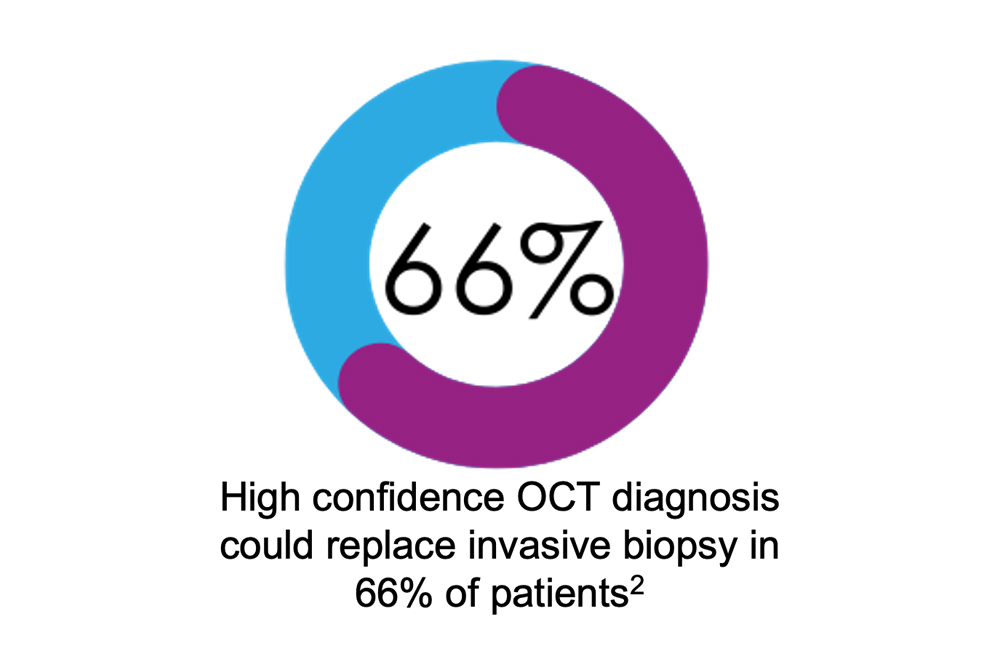
The sensitivity and specificity of OCT for BCC diagnosis have been shown to be non-inferior to invasive biopsy³, with biopsy avoided in up to 66% of patients³.
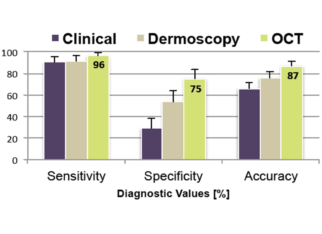
Superior diagnosis compared with traditional non-invasive pathways
Designed for speed, confidence and efficiency
- Full, high‑definition non‑invasive OCT scan in just 15 seconds
- Real‑time placement of AI image markers during acquisition
- Automated lesion penetration depth and volume measurement
- Real‑time 3D volume rendering for rapid lesion visualisation
- Highly cost‑effective — reducing learning curve and examination time
Avoiding biopsy for the majority of BCC patients can simplify the patient pathway and help reduce clinic backlogs.
“The number of biopsies needed are reduced dramatically.”
Prof. U. Paasch, Leipzig, Germany
Reduce biopsy need and speed BCC decision-making
See how VivoSight Dx Pro with VivoAID fits seamlessly into real-world dermatology workflows.
The whole picture — in real time
Only VivoSight Dx Pro provides:
- >1 mm imaging depth for confident treatment selection and BCC characterisation
- Unrivalled 6 × 6 mm field of view for rapid visualisation of the entire lesion
- Real-time 3D OCT volume rendering with optional D-OCT vasculature
- Areas of highest BCC marker density highlighted, accelerating clinician interpretation
Together, these capabilities provide critical context to support confident clinical decision-making.
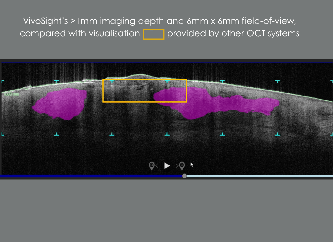
Unrivalled field-of-view and imaging depth, compared with other OCT systems
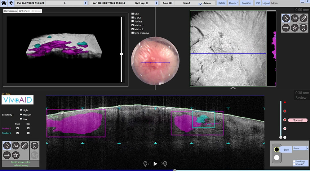
AI-assisted OCT, clinically responsible
VivoAID is designed to support interpretation by highlighting image features during review.
VivoAID supports interpretation by highlighting clinically relevant image features during OCT review—enhancing efficiency while keeping the clinician in control.
- Faster, more consistent workflow with AI-assisted OCT review
- VivoAID acts as a real-time support tool, guiding efficient scan interpretation
- Automatically identifies key BCC features, presented with confidence grading
- Results visualised instantly in 3D, within the native OCT dataset
- Widely adopted by experienced users to optimise clinic workflow efficiency
OCT image analysis and clinical decision-making remain the responsibility of the physician.
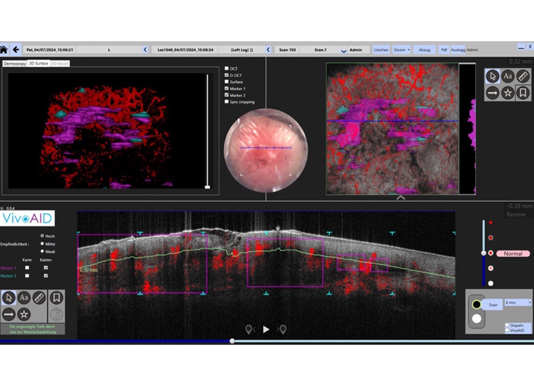
VivoAID image markers and visualisation of microvascular surrounding BCC lesion
“…optical coherence tomography can improve the diagnostic accuracy of difficult-to-recognize BCCs”4
Designed for the clinical dermatologist
VivoAID is purpose-built for the routine dermatology practice—delivering advanced OCT insights without the complexity of research-oriented imaging systems.
Whether you are new to OCT imaging or an experienced user, VivoAID supports faster, more confident adoption. For first-time users, it shortens the learning curve to full clinical integration. For experienced clinicians, the enhanced tools within VivoSight Dx Pro and VivoAID further accelerate interpretation and optimise skin cancer clinic workflows.
Transform NMSC diagnosis with VivoSight
Reduce biopsy. Improve workflow. Strengthen clinical confidence.
From first presentation through treatment selection and follow-up, VivoSight Dx Pro with VivoAID supports faster, more confident and less invasive care for NMSC patients.